Know All About APP Antibody

Amyloid Precursor Protein (APP) or Amyloid-beta precursor protein acts as a cell-surface membrane receptor for kinesin I, responsible for the axonal transfer of beta-secretase and presenilin 1. APP is essential for neuronal growth, neurite adhesion, and the process of axonogenesis. 

The APP protein is a 100-140kDa transmembrane glycoprotein that exists in various isoforms that result from alternative splicing. The proteolytic cleavage of APP through secretases causes the formation beta-amyloid, which is the principal part of plaques that are senile. You can also know more about a beta-Amyloid/APP Antibody online.

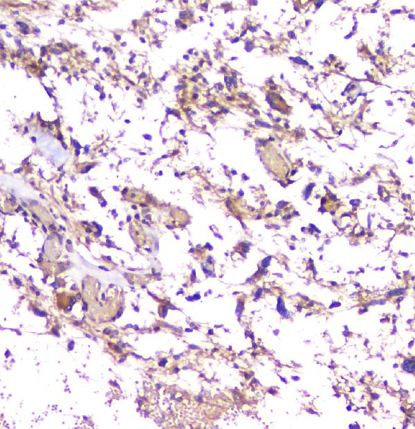
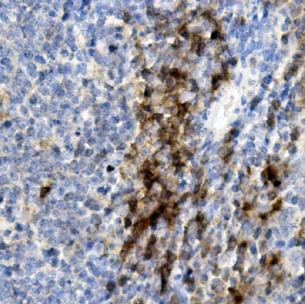
Senile plaques are one the most significant histopathologic manifestations associated with Alzheimer’s. A lack of regulation and processing of the APP is also a factor for Down’s Syndrome, the early onset of familial Alzheimer’s disease and cerebral hemorrhage. This monoclonal antibody recognizes the human mouse and the rat the APP (Amyloid Precursor Protein). 

The APP protein is expressed in high levels within the central nervous system. It comprises three distinct isoforms that result from alternative splicing. APP plays an important role in the formation of synaptic connections and repair and repair, neuronal anterograde transport, iron export and hormonal regulation. 

The secreted APP (sAPP) could be neuroprotective against neurotoxic injury as well as oxidative stress and excitotoxicity. APP is part of a family that includes at most two homologs, amyloid precursors like proteins 2 and 1 (APLP1 and the APLP2). The resemblances in APP and APP and APP, particularly APLP2 suggests that APLP may be able to share and take over the functions of APP. 

What Is CD8 Antibody?

The CD8 beta antibody is an effective marker for cytotoxic lymphocytes. It binds to the CD8 coreceptor on the cell surface and recognizes the topological domain in CD8 alpha. This CD8 antibody recognizes both the CD8 beta heterodimer (the most common), and the CD8 alpha – CD8 b homodimer (the least common). Natural killer cells (NK cells), cortical, and thymocytes also express the CD8A gene.

The CD8A antibody could be used as a marker for these cells. The CD8 molecule is made up of two chains, alpha, and beta. CD8 can be found on T cells of normal cytotoxic/suppressor cells, which account for approximately 20 to 35% of human peripheral blood lymphocytes. You can know more about CD8 antibodies via www.bosterbio.com/anti-cd8-alpha-cd8a-picoband-trade-antibody-a02236-3-boster.html.

Natural killer cells (80% of thymocytes) also detect the CD8 antigen. They are a subpopulation (30% of peripheral blood null cells and 15-30% of bone marrow cells). CD8 + T cells recognize immunogenic peptides present at the cell surface-bound by major histocompatibility class I (MHCI). Antigen recognition is achieved by binding both the T-cell receptor (TCR) and the CD8 co-receptor (pMHCI) to the same peptide -MHCI (MHC). 

The TCR determines the specificity, while CD8 has an effect on antigen sensitivity. To examine the role of CD8 in CD8 + cell activation, anti-CD8 antibodies were extensively used. However, previous studies have shown conflicting results and it is not clear whether anti-CD8 antibody per se can induce effector function. 

OKT8 also increased TCR/pMHCI rates and could therefore be used to enhance pMHCI staining and visualization of antigen-specific T-cells CD8 +. Despite having opposite effects on pMHCI tetramer stained, the anti-mouse CD8 antibody CT-CD8a/CT-CD8b also activated CD8 + T cells. Anti-CD8 antibodies are capable of activating T-cell effector functions in a heterogeneous manner.